Treatment Details
Hello! I'm Dr. Sungmin Ko, the chief director of Seoul Yido Dental Clinic. Since opening the clinic, there's one question I get asked very, very frequently: questions about wisdom teeth.
'Can I get this wisdom tooth extracted here?' Many patients come to our Hongdae wisdom tooth clinic after being told to go to a university hospital. I understand that hearing such advice can be quite alarming.
What are the reasons for being told this? The most common reason is the 'nerve.'
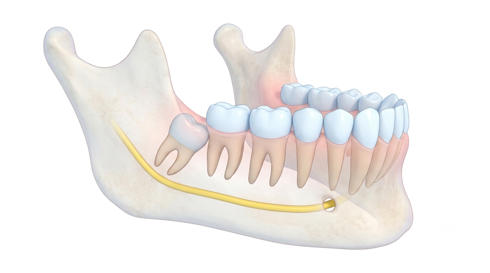
Near the roots of the lower wisdom teeth, there's a nerve called the 'inferior alveolar nerve' that runs through the jawbone. It's a very important nerve responsible for sensation in the lower lip and chin area. If the wisdom tooth root is closely attached to this nerve canal, extra caution is required during extraction,
which is why we sometimes recommend a university hospital.
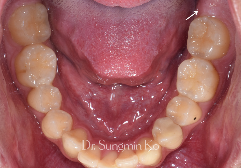
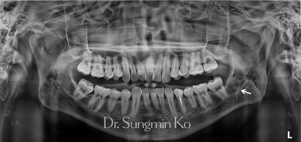
Looking at this photo, the wisdom tooth isn't easily visible inside the mouth, is it? [Image needs confirmation: https://postfiles.pstatic.net/MjAyNjEwMjFfMTYg/MDAxNzkyNTQzOTgzMDAw.Q6lcda0HNv6pBo-2CKjYPByuACIZvCT28RbSTeCLlaUg.9hlu9VEBvbVKcBSzqEQLKyeF4G6EJl8VR_p9jsl4Ysg.PNG/8-3.png?type=w966]
Hongdae Wisdom Tooth Initial Panoramic X-ray: Upon checking the X-ray, it was lying diagonally towards the large molar in front. However, the perceived closeness from a single image can differ from the actual distance.
A panoramic X-ray, commonly taken at dental clinics, is a flat, two-dimensional image of the three-dimensional jawbone. [Image needs confirmation: https://postfiles.pstatic.net/MjAyNjEwMjFfMTQ1/MDAxNzkyNTQ0MDA5NDQz.Q01rOQsaTeLWfPhIeEGro2GgEm_AvRhRnud1tIIbrWQg.NLaMjYaJf64KH_w3AKRnP_iAKR4YlIM239IyToAzeIg.PNG/8-4.png?type=w466]
[Image needs confirmation: https://postfiles.pstatic.net/MjAyNjEwMjFfMjg3/MDAxNzkyNTQ0MDA5MTk5.EqXUqvlLsJlZzi4UoI8hJOPVB2FcWLwcpe4zzChhlrog.k9Ahs3n-7r-Da0NJeyI5tncgl5qaS0Bd1UNc340n9bIg.PNG/8-5.png?type=w466]
Therefore, even if the root and nerve canal appear to overlap in the image, they are often separated in front or behind in reality. It's similar to how two palms held one in front of the other can appear to be touching when a shadow is cast on them.
[Image needs confirmation: https://postfiles.pstatic.net/MjAyNjEwMjFfMjY5/MDAxNzkyNTQ0MDQ5OTI3.SqdRW-wTMSbA0530zzoCUYbcbqOrm945vQa6nUUNwYg.A8M8V3N52iPkXsTTVc7PpePYBSm5Nz7vC3K6KB_itkQg.PNG/8-6.png?type=w966]
In such cases, we first take a CT scan. A CT scan allows us to confirm the 3D position of the root relative to the nerve canal – whether it's in front or behind, and the actual distance.
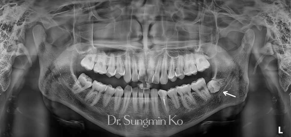
This patient also had an impacted wisdom tooth on the lower left, partially embedded in the gum and bone. However, the CT scan confirmed a safe distance between the root and the nerve canal, allowing for access.
When a wisdom tooth leans against the tooth in front, food can get trapped between them, potentially causing cavities in the front tooth as well, so we recommended extraction. Do impacted wisdom teeth hurt a lot?
If an impacted wisdom tooth is attempted to be removed in one piece, a significant amount of surrounding bone must be removed. Therefore, we divide the tooth into its crown and root sections for removal, which is called sectional extraction.
[Image needs confirmation: https://postfiles.pstatic.net/MjAyNjEwMjFfMTc1/MDAxNzkyNTQ0MDc4MzE4.RiRb3zm-eBnwA2sBbtzKqmat7QhVX8Iv3sGK28XWxSkg.0dKF4KCnVQwm85Tdsk_tzaSSNZpwkc442bRNnDBduf4g.PNG/8-7.png?type=w966]
This is the extracted wisdom tooth after sectional removal. The left is the crown, and the right is the root. By dividing it into pieces, each piece can be removed with less force even in a narrow space, reducing bone removal and minimizing force transmitted to the nerve.
Regarding pain, since the procedure is performed under anesthesia, you won't experience much discomfort during the extraction. However, you might experience some swelling or soreness for a few days afterward!
[Image needs confirmation: https://postfiles.pstatic.net/MjAyNjEwMjFfMzEg/MDAxNzkyNTQ0MTA2OTgy.Xp4QVxUh0WbAl5EUvGJXRyxy1-X2BYzssf6_8jLN2Ywg.HuyJwLBbJJrkJgMPMeVWLo2fFg1t0-N4zZsmAjRvH34g.PNG/8-8.png?type=w966]
This is a post-extraction photo. [Image needs confirmation: https://postfiles.pstatic.net/MjAyNjEwMjFfMTk2/MDAxNzkyNTQ0MTE1NzY0.Keds7APSrnMfMZLuuSZOaSz6pdrouEU2__ywMtF5EjMg.e0Fil3d-t1gWs5GDccD0y82j6c0jg09beQFBKwD6amcg.PNG/8-9.png?type=w466]
[Image needs confirmation: https://postfiles.pstatic.net/MjAyNjEwMjFfMTk2/MDAxNzkyNTQ0MTE1NzI5.XdRzIgzu6YEZTPEEwJVLe4hyg3tkQ9zVf9SD2lQJ6MQg.bspwbnaoko9x8bkNHLB0V3V-VDy_Jqb5VNS_5RmhLqkg.PNG/8-10.png?type=w466]
The space where the wisdom tooth was is now clean and empty, isn't it? The empty space will gradually fill with gum tissue and bone over several weeks to months. Many people tend to take wisdom teeth lightly, thinking they're just teeth to be removed anyway.
I believe that tooth extraction involves not only the removal process but also protecting the adjacent molar and nerve. Therefore, rather than making a judgment based on a single image, I carefully examine it with a CT scan and then discuss the best course of action.
(Of course, if the root surrounds the nerve canal, there's a large cyst around the wisdom tooth, or meticulous care is needed due to a systemic disease, it might be better to proceed at a university hospital.) Wisdom Tooth Extraction, Check with CT First at Hongdae Wisdom Tooth Dental Clinic.
"My wisdom tooth is close to a nerve." "I'm so scared of university hospital extractions." "Should I remove a horizontally impacted wisdom tooth?" If you have these concerns, I recommend accurately checking the condition of your wisdom tooth with a CT scan one more time before deciding on extraction.
Many people are truly afraid of wisdom tooth extraction.

I hope this article was helpful to such individuals! Haha, thank you! This was Dr. Sungmin Ko, Chief Director.
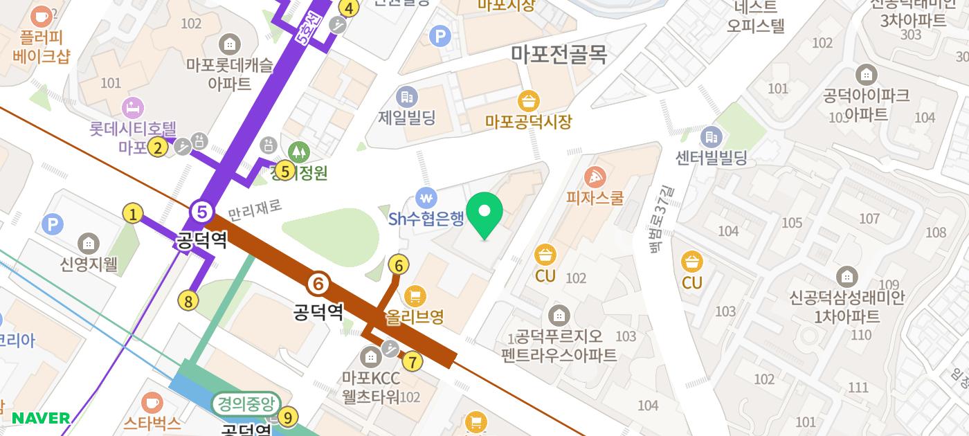
2nd Floor, 24 Mallijae-ro, Mapo-gu, Seoul ※ After wisdom tooth extraction, temporary swelling, pain, bleeding, limited mouth opening, and rarely, sensory abnormalities may occur, requiring regular follow-up observations.